Unveiling Mydriasis: What Dilated Pupils Can Tell You Now
Ever wondered if your eyes are trying to tell you something more than just what you see? Mydriasis, or the persistent and unusual dilation of your pupils, acts as a silent alarm, potentially signaling a range of underlying health issues or external influences that necessitate prompt and thorough evaluation.
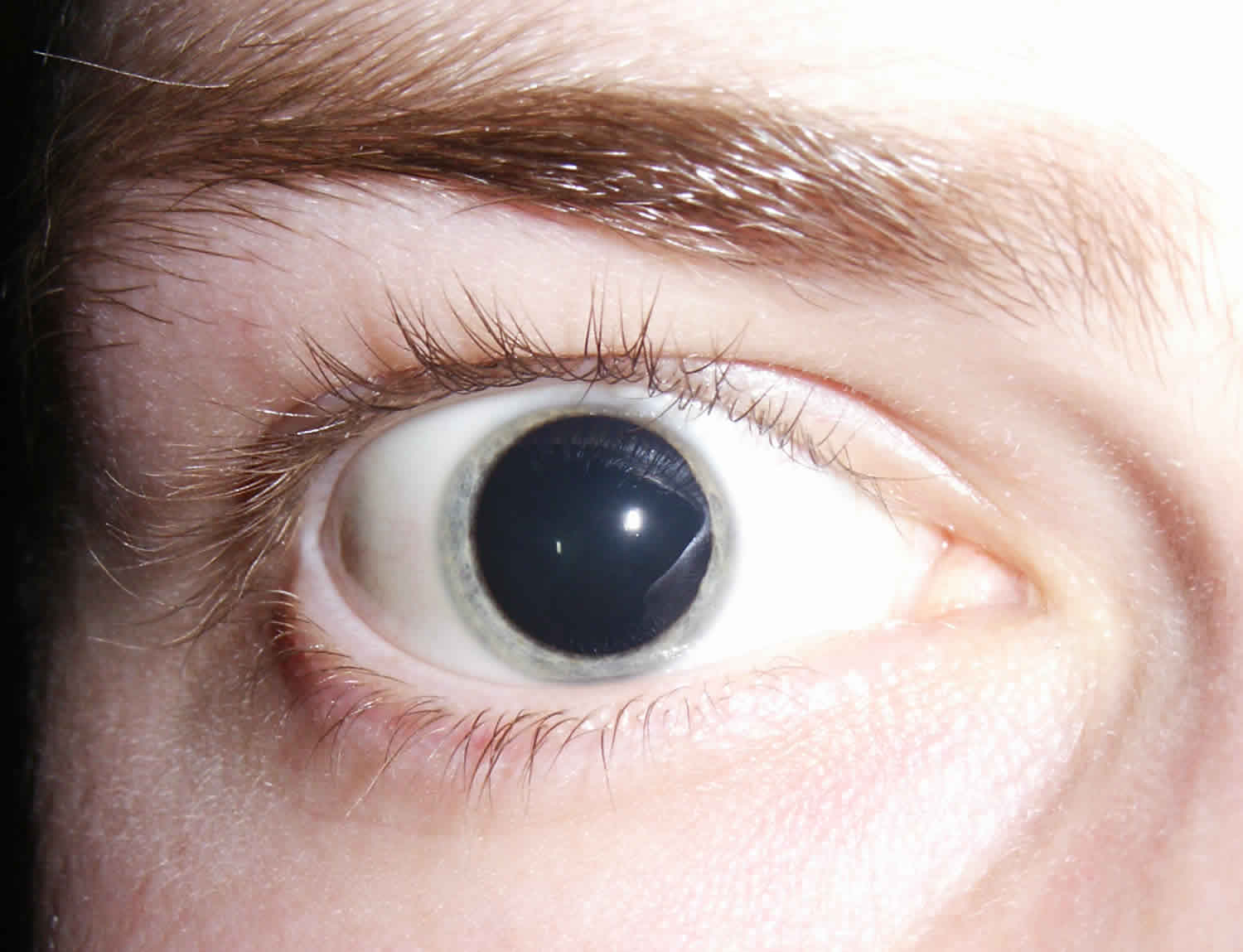
Deep within the iris, that vibrant, colored portion of your eye, lies a delicate muscle responsible for orchestrating the size of your pupil. This intricate dance of expansion and contraction allows your eye to adapt seamlessly to fluctuating light conditions. In dimly lit environments, the pupil gracefully dilates, maximizing light intake, while in brightly illuminated settings, it constricts to prevent overexposure. However, when this finely tuned mechanism falters, resulting in persistent pupil dilation, known as mydriasis, it raises a red flag, suggesting a potential underlying problem.
| Mydriasis: Diagnostic and Clinical Overview | |
|---|---|
| Definition | Abnormal dilation of the pupil, exceeding a diameter of 5 mm, unresponsive to light stimuli or accommodation. |
| Causes |
|
| Symptoms |
|
| Diagnosis |
|
| Treatment |
|
| Complications |
|
| Reference | Mayo Clinic - Dilated pupils |
One of the most significant associations of mydriasis is with angle-closure glaucoma, a serious and potentially vision-threatening eye condition. This is particularly relevant for individuals with what ophthalmologists refer to as "narrow angles," which describes a reduced space between the iris and the cornea. In these individuals, when the pupil dilates, it can obstruct the flow of fluid within the eye, leading to a perilous increase in intraocular pressure. This heightened pressure can damage the optic nerve, potentially resulting in permanent vision loss. Therefore, mydriasis is considered a major risk factor for angle-closure glaucoma in susceptible individuals.
- Phil Hartman The Iconic Comedian Who Left An Indelible Mark
- Unveiling The World Of Xhamsters A Comprehensive Guide
Distinguishing between normal pupil dilation and mydriasis is paramount for timely diagnosis and intervention. Normal pupillary dilation occurs naturally in response to low-light conditions, as the eye attempts to gather more light, or when focusing on distant objects. Conversely, mydriasis is characterized by persistently dilated pupils that exhibit a diminished or absent response to changes in light. This lack of reactivity to light stimuli serves as a crucial diagnostic indicator for identifying mydriasis. It is more commonly observed in individuals predisposed with "narrow angles," which denotes an atypically narrow angle formed between the outer edge of the iris and the cornea, the transparent outer layer that covers the front of the eye.
A myriad of factors can trigger mydriasis, encompassing both internal and external influences. Medications, including both prescription and over-the-counter drugs, can exert a significant impact on pupil size. Certain classes of drugs, such as anticholinergics and antidepressants, are known to induce mydriasis as a side effect. Recreational drugs, particularly stimulants like cocaine and amphetamines, as well as hallucinogens like LSD, are also notorious culprits in causing pupil dilation. Furthermore, physical trauma, especially injuries affecting the eye itself or the brain, can disrupt the normal pupillary response, leading to mydriasis. It's also crucial to be aware of the opposite condition, miosis, characterized by pinpoint pupils, which indicates excessive constriction and may also signal an underlying medical issue.
The importance of assessing pupil size extends far beyond simple visual observation. Elevated pressure within the brain, resulting from conditions such as head injuries, strokes, or tumors, can inflict damage upon the iris muscles, impairing their ability to regulate pupil size effectively. In unconscious patients, the presence of a fixed, dilated pupil warrants immediate and thorough investigation, often raising strong suspicion of a serious intracranial event, such as herniation due to an intracranial mass lesion. Herniation occurs when brain tissue is displaced due to increased pressure, potentially causing irreversible neurological damage. However, it's important to note that a fixed, dilated pupil in an awake and alert patient is typically not indicative of herniation and points towards other possible etiologies.
- Unveiling The Truth About Xhamster A Comprehensive Guide
- Unlock Your Future Your Ultimate Guide To Degree Navigator Rutgers
Mydriasis can also arise as a consequence of the dilating eye drops administered during routine eye health examinations. These eye drops temporarily widen the pupils, enabling the ophthalmologist to gain a more comprehensive view of the back of the eye, including the retina, optic nerve, and blood vessels. This induced dilation is a controlled and transient effect, and it differs significantly from the mydriasis caused by underlying medical conditions or drug exposure. The effects of these eye drops usually subside within a few hours, restoring the pupils to their normal size.
Mydriasis, with its noticeable alteration in eye appearance, often attracts attention. However, the causes of this pupillary dilation are diverse and require careful evaluation. Several ocular disorders can disrupt the normal mechanisms that regulate pupil constriction. Diagnosing the underlying cause of mydriasis typically involves a multi-faceted approach. The etiologies are broad, encompassing a spectrum of medical conditions to medication side effects, each necessitating tailored and specific treatment strategies.
Determining whether pharmacologic mydriasis, induced by topical agents, is present necessitates a thorough examination and evaluation. Certain eye drops, employed for diagnostic or therapeutic purposes, have the potential to cause pupillary dilation. A meticulous review of the patient's medication history, coupled with a comprehensive eye examination and potentially specific diagnostic testing, can help pinpoint the underlying cause of the mydriasis.
The remarkable capacity of the eye for recovery is exemplified by a case involving traumatic mydriasis and accommodative dysfunction resulting from a sweetgum ball ocular injury, with full resolution observed eight years following the initial trauma. This underscores the significance of long-term monitoring and highlights the body's intrinsic regenerative capabilities.
The counterpart to mydriasis is miosis, characterized by pinpoint pupils, where the pupils exhibit abnormal constriction. Both of these pupillary abnormalities can serve as symptomatic indicators of an underlying health condition, warranting prompt medical evaluation. In the German language, the dilation of the pupil exceeding 5mm in diameter is expressed as "Als Mydriasis bezeichnet man die Weitstellung der Pupille ber einen Durchmesser von 5 mm hinaus."
The application of a comprehensive ophthalmology system like MYDRSI provides a structured and systematic methodology for diagnosis and management of various eye conditions. It provides standardized diagnostic protocols, personalized treatment plans, seamless technological integration, and meticulous monitoring capabilities. Essentially, MYDRSI empowers practices to manage patient records, schedule appointments, and streamline insurance claim processing with heightened efficiency.
Mydriasis occurs when your pupils dilate unnaturally, independently of ambient light levels. This atypical dilation, uncoupled from environmental lighting cues, serves as a crucial identifier and sparks a multitude of diagnostic inquiries.
Joining a program or utilizing a digital platform provides an array of benefits, including complimentary enrollment, access to comprehensive course materials, and valuable certifications. Additional advantages include the ability to interact and collaborate with other users and effectively manage one's professional online presence.
The differential diagnosis for mydriasis is broad, spanning from benign physiological responses to serious neurological emergencies. It necessitates careful consideration of the patients history, medication list, and associated symptoms to arrive at an accurate diagnosis. Physiological mydriasis can occur in response to fear, anxiety, pain, or sexual arousal. Pathological mydriasis may indicate underlying conditions such as third cranial nerve palsy, Horners syndrome, Adies tonic pupil, or drug toxicity.
Imaging studies, such as computed tomography (CT) or magnetic resonance imaging (MRI) of the brain, may be warranted to evaluate for intracranial lesions, tumors, or vascular abnormalities that can compress or disrupt the pupillary pathways. Pharmacological testing, involving the administration of pilocarpine eye drops, can help differentiate between various causes of mydriasis based on the pupillary response. In cases of suspected angle-closure glaucoma, gonioscopy is essential to assess the drainage angle and determine the risk of acute angle closure.
The management of mydriasis is directed at addressing the underlying cause. If mydriasis is attributed to medications, discontinuation or substitution of the offending agent may be necessary. In cases of angle-closure glaucoma, urgent intervention is required to lower intraocular pressure and prevent irreversible optic nerve damage. This may involve pharmacological agents, such as miotics and topical or systemic pressure-lowering medications, as well as surgical procedures, such as laser iridotomy or peripheral iridectomy.
Neurological causes of mydriasis, such as third cranial nerve palsy, may necessitate further investigation to identify the underlying etiology, which can range from aneurysms and tumors to inflammatory or infectious processes. Treatment may involve medical management, surgical intervention, or a combination of both, depending on the specific diagnosis. In cases of traumatic mydriasis, observation may be sufficient if there is no underlying structural damage. However, if the pupil remains persistently dilated and symptomatic, surgical repair may be considered to improve cosmesis and reduce glare or photophobia.
Complications of mydriasis can include blurred vision, particularly at near, sensitivity to light (photophobia), and glare. These symptoms can significantly impact visual function and quality of life. In cases of angle-closure glaucoma, the most serious complication is irreversible vision loss secondary to optic nerve damage. Neurological causes of mydriasis can lead to a wide range of neurological deficits, depending on the underlying condition.
The prognosis for mydriasis depends on the underlying cause and the promptness of diagnosis and treatment. In cases of medication-induced mydriasis, the pupil typically returns to normal size upon discontinuation of the offending agent. Angle-closure glaucoma requires timely intervention to prevent vision loss, and the prognosis is generally good if treated promptly. Neurological causes of mydriasis can have variable prognoses, depending on the underlying condition and the effectiveness of treatment.
Education and counseling are essential components of managing mydriasis. Patients should be educated about the potential causes of mydriasis, the importance of seeking prompt medical attention, and the potential complications associated with the condition. They should also be advised on strategies to minimize symptoms, such as wearing sunglasses to reduce photophobia and glare.
Regular follow-up with an ophthalmologist or neurologist is crucial to monitor the condition and ensure that appropriate treatment is initiated and maintained. Patients should be instructed to report any changes in vision, eye pain, headaches, or other neurological symptoms to their healthcare provider.
The accurate diagnosis and management of mydriasis require a multidisciplinary approach involving ophthalmologists, neurologists, and other healthcare professionals. Collaboration and communication among these specialists are essential to ensure that patients receive comprehensive and coordinated care.
Ongoing research is aimed at improving the understanding of the underlying mechanisms of mydriasis and developing new and more effective treatments for the condition. Areas of investigation include the development of novel pharmacological agents to modulate pupillary size, advanced imaging techniques to visualize the pupillary pathways, and innovative surgical approaches to repair damaged iris muscles.
The information provided herein is intended for general knowledge and informational purposes only, and does not constitute medical advice. It is essential to consult with a qualified healthcare professional for any health concerns or before making any decisions related to your health or treatment.
A thorough examination of the pupillary response is a cornerstone of the neurological examination. The size, shape, and reactivity of the pupils to light provide valuable clues about the integrity of the brainstem and cranial nerves. Asymmetry in pupil size (anisocoria) or abnormal pupillary reflexes can indicate underlying neurological pathology.
The pupillary light reflex is a fundamental neurological pathway that involves the retina, optic nerve, optic chiasm, pretectal nucleus, Edinger-Westphal nucleus, and ciliary ganglion. Afferent signals from the retina travel along the optic nerve to the pretectal nucleus in the midbrain, which then projects bilaterally to the Edinger-Westphal nuclei. Efferent signals from the Edinger-Westphal nuclei travel along the oculomotor nerve (third cranial nerve) to the ciliary ganglion, which innervates the pupillary sphincter muscle, causing pupillary constriction.
Damage or disruption to any part of this pathway can result in abnormal pupillary reflexes, including mydriasis or miosis. For example, a lesion of the optic nerve can cause an afferent pupillary defect (APD), characterized by decreased pupillary constriction in response to light shined in the affected eye. A lesion of the oculomotor nerve can cause mydriasis due to paralysis of the pupillary sphincter muscle.
Certain medications, such as anticholinergics, can block the action of acetylcholine at the pupillary sphincter muscle, leading to mydriasis. These medications are commonly used to treat a variety of conditions, including overactive bladder, irritable bowel syndrome, and Parkinson's disease. Recreational drugs, such as cocaine and amphetamines, can also cause mydriasis by stimulating the sympathetic nervous system.
Eye injuries, such as blunt trauma or penetrating injuries, can damage the iris or pupillary sphincter muscle, leading to mydriasis. In some cases, the damage may be permanent, resulting in a fixed, dilated pupil. Neurological disorders, such as Horner's syndrome and third nerve palsy, can also cause mydriasis. Horner's syndrome is characterized by miosis, ptosis (drooping eyelid), and anhidrosis (decreased sweating) on the affected side of the face, while third nerve palsy is characterized by mydriasis, ptosis, and ophthalmoplegia (paralysis of eye muscles).
Mydriasis can also be a sign of increased intracranial pressure, which can occur in conditions such as head injury, stroke, or brain tumor. The increased pressure can compress the oculomotor nerve, leading to mydriasis. In these cases, mydriasis is a late sign and may indicate impending herniation of the brainstem.
The evaluation of mydriasis typically involves a thorough history and physical examination, including a detailed neurological examination. The history should include questions about medications, drug use, eye injuries, and neurological symptoms. The physical examination should include assessment of visual acuity, pupillary reflexes, eye movements, and cranial nerves.
The use of magnifying devices like slit lamps allows doctors to see structures in greater detail. It helps in accurate diagnosis of the reason behind the dilation of pupils. It helps to identify the problem, so treatment is done accurately.
While dilated pupils might seem like a minor detail, they can be an indicator of a larger issue, and could be a sign of head trauma, eye injury, reactions to a medication or drug, or any number of underlying medical conditions.
- Unveiling The Xhm Craze A Deep Dive Into The Phenomenon Taking Over The World
- Mike Faist The Rising Star Whorsquos Stealing Hearts With His Talented Performances
.jpg)
What Is Miosis of the Eye and How to Manage It OBN
Mydriasis, mydriatic pupil causes, diagnosis & treatment

Mydriasis What Is It, Pronunciation, Causes, Glaucoma Risk Factor, and More Osmosis
